Abstract
SHS investigation development is considered from the geographical and historical viewpoint. 3 stages are described. Within Stage 1 the work was carried out in the Department of the Institute of Chemical Physics in Chernogolovka where the scientific discovery had been made. At Stage 2 the interest to SHS arose in different cities and towns of the former USSR. Within Stage 3 SHS entered the international scene. Now SHS processes and products are being studied in more than 50 countries.


Abstract
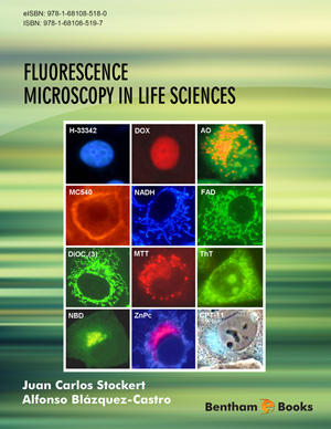
The visualization of cell structures by fluorescent vital probes to study cell organelles, cell viability and signaling processes has great importance in biomedical sciences. Main interest is devoted to the uptake, accumulation and prediction of localization of drugs and xenobiotics in cultured cells. Mechanistic aspects and predictive rules are studied using numerical parameters, which conform the quantitative structure-activity relationships (QSAR) for vital probes. The uptake of probes into cells is the first step for labeling. Several mechanisms of cell uptake and accumulation in cell organelles are known, the most relevant being passive diffusion and active endocytosis (i.e. adsorptive and fluid-phase, binding to receptors). In addition to theoretical predictions, direct cytotoxic effects, photodynamic action, and fading must be taken into account in the microscopical evaluation of uptake and localization of fluorescent probes.
Keywords:
Acridine orange, Amphiphilicity, Autofluorescence, Benzothiazoles, Chromatin DNA, Colocalization, DiOC1(3), Golgi apparatus, Hoechst dyes, Hydroethidine, Hydrophobic index, Log P, Lysosomes, Merocyanine 540, Mitochondria, Nile red, QSAR, Rhodamine 123, Trypan blue.
Recommended Chapters
We recommend

Authors:Bentham Science Books